


Abstract
The aim of this study was to test the hypothesis that neural respiratory drive, measured using diaphragm electromyogram (EMGdi) activity expressed as a percentage of maximum (EMGdi%max), is closely related to breathlessness in chronic obstructive pulmonary disease. We also investigated whether neuroventilatory uncoupling contributes significantly to breathlessness intensity over an awareness of levels of neural respiratory drive alone.
EMGdi and ventilation were measured continuously during incremental cycle and treadmill exercise in 12 chronic obstructive pulmonary disease patients (forced expiratory volume in 1 s±sd was 38.7±14.5 % pred). EMGdi was expressed both as EMGdi%max and relative to tidal volume expressed as a percentage of predicted vital capacity to quantify neuroventilatory uncoupling.
EMGdi%max was closely related to Borg breathlessness in both cycle (r=0.98, p=0.0001) and treadmill exercise (r=0.94, p=0.005), this relationship being similar to that between neuroventilatory uncoupling and breathlessness (cycling r=0.94, p=0.005; treadmill r=0.91, p=0.01). The relationship between breathlessness and ventilation was poor when expansion of tidal volume became limited.
In chronic obstructive pulmonary disease the intensity of exertional breathlessness is closely related to EMGdi%max. These data suggest that breathlessness in chronic obstructive pulmonary disease can be largely explained by an awareness of levels of neural respiratory drive, rather than the degree of neuroventilatory uncoupling. EMGdi%max could provide a useful physiological biomarker for breathlessness in chronic obstructive pulmonary disease.
Abstract
Exertional breathlessness in patients with severe COPD is closely related to levels of neural respiratory drive http://ow.ly/BO6MI
Introduction
Breathlessness is an important cause of exercise limitation and reduced quality of life for patients with chronic obstructive pulmonary disease (COPD) [1, 2]. Our understanding of the psychophysiological mechanisms underlying breathlessness remains incomplete [2, 3]. Although it is widely appreciated that an awareness of levels of motor output to the respiratory muscles from the brainstem respiratory centre is important to the sensation of breathlessness 4, the identification of reliable physiological measures of neural respiratory drive (NRD) poses a significant challenge. Ventilation does not adequately reflect NRD in COPD because changes in the mechanical properties of the respiratory system alter the relationship between NRD and inspiratory flow [5]. Similarly, since respiratory muscle pressure generation is dependent on the contractile function of the respiratory muscles and the length-tension properties of the muscle, independent the NRD level, pressure-derived variables underestimate NRD when pulmonary hyperinflation is present [5, 6].
Breathlessness has also been shown to increase disproportionally if the ventilatory response is limited by impaired pulmonary mechanics (neuroventilatory uncoupling (NVU)) [7]. To date, the relationship between NVU and breathlessness in COPD has been most extensively studied using the “effort/displacement ratio” (EDR). The EDR is the ratio of tidal swings of oesophageal pressure (Poes) relative to maximum inspiratory pressure (PImax) i.e. “effort” and the tidal volume (VT) response expressed relative to the predicted vital capacity (VC) i.e. “displacement”, thus giving the ratio of Poes/PImax to VT/predicted VC [8]. The EDR correlates well with Borg breathlessness in normal subjects and in patients with chronic airflow limitation [8], increasing markedly with breathlessness after a critically low inspiratory reserve volume (IRV) has been reached [9]. However because the oesophageal pressure swing is reduced by hyperinflation [10], the reliability of indices such as the EDR, which rely on pressure-derived measures of NRD, is also limited for COPD.
Recent advances in measuring the diaphragm electromyogram (EMGdi), using multi-pair oesophageal recording electrodes by our group [11–14] and others [15–18], have enabled the accurate quantification of NRD and NVU in terms of a measurement that is neurophysiologically more “upstream” of respiratory pressure generation. The aim of the present study was to investigate the relationship between NRD and breathlessness, using EMGdi as an index of NRD. The influence of NVU on increases in the intensity of breathlessness was also assessed, using the ratio of EMGdi activity to ventilatory output as an index of NVU. These relationships were examined during maximal cycle and treadmill exercise tests to assess the consistency of relationships during different forms of exercise.
Methods
Patients
12 COPD patients (mean±sd) age 66.7±7.0 years, forced expiratory volume in 1 s (FEV1) 38.7±14.5 % pred, n=11 males) were studied. Participants were recruited in accordance with local research ethics committee procedures, and written informed consent was given. Age, height, weight, body mass index and Medical Research Council dyspnoea score were documented.
Participants made two visits at least 24 h apart. FEV1 and slow VC were measured on both days. Inspiratory capacity (IC) and lung volumes (using body plethysmography) were measured prior to exercise on visit 2 only.
Exercise protocols
Visit 1
Incremental cycle and incremental treadmill exercise tests were undertaken to familiarise participants with the exercise and breathlessness assessment protocols. The patients were asked to score breathlessness intensity (strength of sensation) and the intensity of leg fatigue using the modified Borg scale [19]. Each subject was briefed by explaining that descriptors of the intensity of the sensations on the Borg scale are anchored to numerical points on the scale, e.g. for breathlessness, varying between 0 (no breathlessness) and 10 (maximum breathlessness the patient had ever experienced). Further details of the equipment and exercise protocols are provided in the online data supplement. Modified Borg breathlessness and leg fatigue scores were assessed at baseline, at the end of each minute of exercise and at the time of exercise limitation by symptoms (“symptom limitation”) in both protocols.
Visit 2
EMGdi was recorded using a multi-pair oesophageal catheter [20] and ventilation recorded using a pneumotachograph connected to the patient via the full face mask with a noseclip in place. Following measurements made seated at rest, subjects performed the incremental cycle and treadmill exercise tests to symptom limitation, with Borg breathlessness and leg fatigue assessments as in visit 1. At recruitment, patients were alternately assigned to undertake either the cycle or treadmill exercise first. Both exercise tests were performed on the same day with a minimum rest of 1 h between. Oxygen uptake, carbon dioxide production and minute ventilation (V′E) were measured throughout exercise (AD Instruments, Castle Hill, Australia), using a full face mask. Data were acquired using a desktop computer (Apple Computer Inc, Cupertino, CA, USA) running Chart software (version 5.4.2, AD Instruments). Metabolic data were analysed online and stored for further offline analysis. Haemoglobin oxygen saturation was assessed by pulse oximetry in all patients. IC was assessed immediately before each exercise test and as close to the exercise termination point as possible.
Data analysis
EMGdi, metabolic and other cardiorespiratory data were averaged over 30-s time periods. Group data were reported at each 20% epoch of exercise time (i.e. from the end of the rest period onwards) and the patterns of increase in each variable during each exercise modality examined.
EMGdi activity, expressed as a percentage of maximum (EMGdi%max), was calculated per breath as the mean peak root mean square (RMS) EMGdi per breath expressed as a percentage of the peak RMS EMGdi activity, which were recorded during inspiratory manoeuvres performed before exercise (inspiration to total lung capacity from functional residual capacity (FRC), maximum sniff from FRC, PImax manoeuvre against a closed shutter from FRC and sprint maximum voluntary ventilation >15 s). In addition EMGdi%max was also calculated during performance of IC manoeuvres at end exercise, and during breaths taken throughout the exercise protocols. The NVU index was calculated as the ratio of EMGdi%max to VT expressed as a percentage of predicted vital capacity i.e. EMGdi%max/VT%VCpred). Further details, including EMGdi signal processing and analysis of composite indices derived from EMGdi%max, and respiratory rate and inspiratory time, are provided in the online supplementary material.
Analysis of slopes of the relationships between Borg breathlessness, EMGdi%max and ventilation after the “breathlessness threshold”
Previous studies in both healthy subjects and patients have shown that patterns of increases in breathlessness usually follow an alinear “tick” shape, with little change in breathlessness over the first few minutes of exercise, followed by a phase of rapid increases in breathlessness after the breathlessness threshold [9, 14]. The breathlessness threshold was determined by plotting individual Borg breathlessness time-curves throughout exercise, identifying, by eye, a point of inflection for each subject as the point of intersection of two linear relationships. In the present study, for each patient, each physiological variable was normalised to the value of that variable at the timepoint at which the slope of the breathlessness–time relationship became more steeply positive at the breathlessness threshold (figure S1). For example, if V′E at baseline, 20% and 40% epochs of exercise time were 10, 20 and 25 L·min−1, respectively, and breathlessness increased sharply at 20% exercise time, the normalised values of V′E were 10/20 (0.5), 20/20 (1) and 25/20 (1.25) at baseline, 20% and 40% exercise time, respectively, a normalised value of 2 indicating doubling of that variable relative to the take-off point. This allowed the slopes of the increases in each variable to be compared regardless of the absolute numerical value. The purpose of this process was to evaluate the relationships between incremental changes in physiological variables with increases in breathlessness, so as to evaluate the discriminatory powers of each variable. More positive (“steeper”) slopes would indicate that increases in breathlessness were associated with little incremental numerical increase in the physiological variable, indicating that changes in such variables discriminated poorly between successive step increases in the intensity of breathlessness.
Statistical analysis
Statistical analysis was carried out using SPSS software version 15.0.1 for Windows (SPSS Inc, Chicago, IL, USA) and Graphpad Prism 5 for Windows v5.00 (Graphpad Software Inc, La Jolla, CA, USA). EMGdi%max was non-normally distributed during exercise and, therefore, described as median (interquartile range (IQR)) and analysed using nonparametric statistics. Inter-subject comparisons were made using Wilcoxon signed-rank tests (nonparametric) unless otherwise stated. A p-value <0.05 was considered statistically significant.
Results
The results obtained during cycle and treadmill exercise testing, were similar. Consequently only the results from the cycle exercise are reported here to highlight the principal findings. The treadmill data and cycle/treadmill comparisons are reported in the online supplementary data. Results of analysis for composite indices derived from EMGdi%max, respiratory rate and inspiratory time are also reported in the online supplementary data.
Demographic and anthropometric data, spirometry, and lung volume measurements are shown in table 1. Lung volume measurements were obtained from only eight patients.
Symptoms, ventilation, metabolic data and EMGdi activity before exercise and at the point of symptom limitation during cycle exercise are shown in table 2. When cycling, eight patients stopped because of breathlessness, two stopped because of leg fatigue and a further two stopped because of both breathlessness and leg fatigue. When on the treadmill, seven patients stopped because of breathlessness, three patients stopped because of leg fatigue and a further two patients stopped because of both breathlessness and leg fatigue.
Relationships between EMGdi%max, ventilation, NVU and breathlessness during cycle exercise
A positive inflection point, the breathlessness threshold was observed at median (IQR) of 30% (20–40%) exercise time, above which there was a near-linear increase in breathlessness from the low levels observed early in exercise (fig. 1 and S1). EMGdi%max increased in parallel with increases in breathlessness (fig. 2). This was in contrast to the pattern of increase observed in VT%VCpred, which initially increased linearly but reached a plateau at ∼40% of cycle-exercise time (fig. 2).

Increases in Borg breathlessness during 20% epochs of cycle-exercise time. Data points represent median and interquartile range.
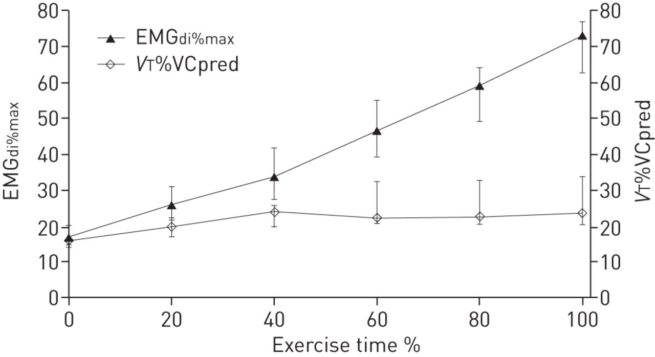
Tidal volume expressed as a percentage of predicted vital capacity (VT%VCpred) and mean root mean square (RMS) diaphragm electromyogram (EMGdi) activity per breath, expressed as a percentage of maximum RMS EMGdi recorded during maximal inspiratory manoeuvres (EMGdi%max) during 20% epochs of cycle-exercise time. Data points represent median and interquartile range.
Median (IQR) correlation coefficients, obtained from individual patients, indicated breathlessness was closely related to EMGdi%max during cycle exercise (r=0.98 (0.94–0.99), p=0.002) (fig. 2, table 3). Despite compensatory increases in respiratory rate, a plateau in ventilation, defined <10% increase in V′E between successive 20% time epochs accompanied by a >10% increase in EMGdi%max, occurred in six out of 12 patients during cycle exercise between 60% and 80% exercise time. This resulted in breathlessness increasing despite very little incremental change in V′E. By contrast, EMGdi activity continued to increase, leading to better discrimination between successive Borg scores using EMGdi%max (fig. 3). After the onset of NVU, increases in breathlessness were accompanied by an increases in the EMGdi%max median (IQR) slope 3.76 (2.04–4.80), but not by an increases in V′E slope 0.31 (−0.06–0.78), the difference between slopes for the EMGdi%max breathlessness and V′E breathlessness relationships was statistically significant (p=0.03). The correlation between NVU (EMGdi%max/VT %VCpred) and breathlessness was r=0.94 (0.84–0.99) (p=0.005) (table 3) and statistically no stronger than that between EMGdi%max and breathlessness.

A comparison of the relationship between minute ventilation (V′E), breathlessness and the mean root mean square (RMS) diaphragm electromyogram (EMGdi) activity per breath, expressed as a percentage of maximum RMS EMGdi recorded during maximal inspiratory manoeuvres (EMGdi%max) during 20% epochs of cycle-exercise time. V′E and EMGdi%max have been plotted on the same scale to highlight the difference in the slopes of the relationships with breathlessness after the onset of neuroventilatory uncoupling. Data points represent median and interquartile range.
EMGdi%max, ventilation and breathlessness relationships during cycle exercise after the breathlessness threshold
For each patient, physiological variables were normalised to the value of the variable at the breathlessness threshold (fig. S1). Every normalised variable except VT%VCpred, demonstrated a significantly greater slope of its relationship with breathlessness after the breathless threshold than before (table 4). After the breathlessness threshold, incremental changes in EMGdi%max were significantly greater and less variable than incremental changes in ventilation as the intensity of breathlessness increased, as indicated by the slope of the normalised EMGdi%max and breathlessness relationship being significantly (p=0.0005) less than the slope of the normalised V′E and breathlessness relationships (EMGdi%max 5.7 (3.0–6.6) versus V′E 10.4 (7.0–19.8)).
Discussion
The principal finding of this study is that neural respiratory drive, quantified as EMGdi%max is closely related to exertional breathlessness in COPD. Increases in EMGdi%max discriminates better between successive Borg breathlessness scores than increases in ventilation, which plateaued as a consequence of significant NVU due to impaired pulmonary mechanics.
These data support the hypothesis that conscious awareness of respiratory motor output is important to the perception of breathlessness in COPD. This is in keeping with previous findings by Duiverman et al. [21], who showed increased intercostal and scalene EMG activity to be associated with increased breathlessness in healthy subjects and in COPD patients. Diaphragm and parasternal intercostal muscle EMG have also been recently shown to closely correlate with breathlessness in cystic fibrosis, before and after the onset of neuromechanical dissociation when the ventilation–breathlessness relationship again became less strong [14]. A close relationship between breathlessness and the EMG of the parasternal intercostals, scalene, and alae nasi muscles, has also been observed in mechanically-ventilated intensive care patients [22]. Similar to the results presented here, the strength of the relationship to breathlessness in the study by Schmidt et al. [22] was not improved (and in some cases deteriorated) when ventilation achieved in response to increases in NRD was considered. Also, in healthy young males and females [23, 24], and in the altered “normal” physiological states of advancing age, obesity, and pregnancy [25–27], breathlessness can be mostly explained by an increased awareness of NRD (measured as V′E and ΔPoes/PImax) with little contribution by impaired respiratory mechanics, particularly at submaximal exercise. Our data suggest that in COPD, NVU similarly contributes little to the sensation of breathlessness over an awareness of the levels of increased NRD alone. The correlation between EMGdi%max/VT%VCpred and breathlessness was at best numerically similar to that between EMGdi%max and breathlessness. This is out of keeping with the view that length-tension inappropriateness drives breathlessness [28], a hypothesis supported by the observation recently made by O’Donnell et al. [9] that breathlessness increased when IRV fell below a threshold level, accompanied by a sharp increase in the EDR. Methodological differences are important here. Unlike EMGdi, respiratory-muscle pressure generation will underestimate NRD in COPD because there is significant neuromechanical uncoupling. The increase in breathlessness at the dyspnoea-IRV inflection point, identified in the study by O’Donnell et al. [9], was in fact less steep than an increase in breathlessness observed on inspection of their data from an earlier stage in exercise. This earlier timepoint was not associated with an increase in the EDR, but associated increases in NRD could have been evident using EMGdi.
In the present study EMGdi%max was 73.3% at end-cycle exercise and 69.9% at end-treadmill exercise, which were consistent with previously reported levels [13, 16]. It is possible that this level of activation represents a submaximal threshold above which further diaphragm activation does not lead to useful increases in tension generation or ventilation [16]. It was interesting to note that most patients reached EMGdi%max values in excess of 25%, and in some cases beyond 40%, before the breathlessness threshold was reached. These levels of EMGdi%max are greater than those observed at an equivalent point in our study investigating the relationship between EMGdi%max and breathlessness in cystic fibrosis patients (19%) and healthy subjects (9%) [14]. This indicates that patients with COPD, whose levels of NRD are already higher than those of healthy individuals [12, 15], tolerate higher levels of NRD than would usually be dyspnegenic in healthy subjects, i.e. the perception of breathlessness associated with high levels of NRD appears to be “blunted” in severe COPD. This is consistent with the common clinical observation that patients with chronic lung disease often tolerate high mechanical respiratory loads without reporting significant breathlessness. The COPD patients in this study were able to tolerate a higher level of EMGdi%max before feeling breathless when compared to the cystic fibrosis patients in our previous work [14]. This is in keeping with “desensitisation” to a mechanical load on the respiratory system, which is higher and is present for a longer duration in COPD in comparison cystic fibrosis. Reorganisation of the sensory cortex in COPD is certainly plausible given that we have shown changes in corticospinal pathway excitability in this condition [29, 30]. In asthma, a low baseline FEV1 and high bronchial responsiveness have been shown to be associated with a low degree of “perceptiveness” of bronchoconstriction [31]. This desensitisation might also reflect the shift from type II towards type I fibres in COPD diaphragm muscle, which is regarded to confer the beneficial effect of rendering the COPD diaphragm more resistant to fatigue [32].
Limitations of the study and techniques used
IC was not measured during exercise and, therefore, it was not possible to examine the association between changes in EMG activity and dynamic hyperinflation. The relationship between NRD, neuromechanical uncoupling and breathlessness around critically-low IRVs, implicated in the acceleration of exertional breathlessness to intolerable levels in COPD [9], could be the focus of future studies, shedding light on the events critical to the breathlessness threshold and the point at which patients stop exercise. In the present study, NVU was evident from the exercise onset during both forms of exercise but more pronounced over the latter 40–60% of exercise time which corresponded to the limit of expansion of VT during exercise (fig. 2). Whereas O'Donnell et al. [9] found that the threshold of dyspnoea corresponded to the time of limited expansion of VT, associated with a critical threshold of IRV, the breathlessness threshold in the present study occurred before the onset of VT limitation and was related to increases in EMGdi%max. Although identification of the breathlessness threshold in the present study was by eye, the point of intersection for the breathlessness time lines was in general quite clear and, therefore, any error is likely to be minimal.
All of the patients participating in this study tolerated the oesophageal catheters well, but it is appreciated that there are other noninvasive methods for recording respiratory muscle EMG activity that could have given similar results. However, we believe that there are distinct advantages to using oesophageal multi-pair recording electrodes to quantify the diaphragm EMG, particularly when the respiratory system is fully loaded during exercise in a severe COPD patient group. First, the diaphragm is the principal inspiratory muscle, accounting for ∼70% of V′E in normal humans [33] and the majority of inspiratory work [34]. It is acknowledged that despite this, earlier work in healthy subjects has suggested that the sensation of respiratory effort in nonexercise tasks is related to an awareness of increased NRD directed to extradiaphragmatic obligate inspiratory muscles and accessory muscles of inspiration, rather than NRD to the diaphragm itself [35, 36]. Quantification of NRD using surface recordings of another obligate inspiratory muscle, parasternal intercostal muscle EMG activity (EMGpara), have the advantage over EMGdi recorded using oesophageal catheters of being noninvasive. Surface recordings of EMGpara have also been shown to be closely related to breathlessness in healthy subjects [14, 21] and in respiratory disease [14, 21, 22, 37]. However, work by our group [13, 14, 20] and others [16, 24] has shown that EMGdi activity in patients with chronic airflow limitation and healthy individuals increases continuously throughout the duration of incremental exercise protocols, and that breathlessness is closely related to EMGdi activity in healthy subjects [14, 24] and in cystic fibrosis [14]. In COPD, improvements in breathlessness following lung volume reduction surgery are closely related to reductions in NRD as assessed by EMGdi [38]. These results are in contrast to earlier findings, and are in keeping with the hypothesis that breathlessness in COPD patients is closely related to an awareness of motor drive to the diaphragm, rather than an awareness of NRD to extradiaphragmatic muscles alone. One explanation for the difference in the findings is likely to be the significant advances in EMGdi recording technology and analysis software that have been made over recent decades. This includes the development of oesophageal catheters wired as multiple, overlapping, pairs of bipolar electrodes and analysed using digital analysis algorithms [12–16, 18, 20, 24, 39, 40], which have greatly improved the reliability and signal-to-noise ratio of the technique over earlier methods [35, 36, 41, 42].
Secondly, although the parasternal intercostal muscles are activated synchronously with the diaphragm at rest, the pattern of recruitment of the diaphragm and parasternal intercostal muscles, as NRD increases, is similar but not identical. As shown by Gandevia et al. [43], the parasternal intercostal muscles and diaphragm adopt different “strategies” to increase motor unit output when NRD increases; the diaphragm showing a predominance for frequency modulation, and the parasternal intercostal muscles a predominance for motor unit recruitment. Furthermore, diaphragm activation approaching the upper limit of ventilatory capacity appears to be closer to maximum than that of the parasternal intercostal muscles under the same condition. Whereas EMGdi%max reaches near-maximal levels at the end of exercise, surface EMGpara%max levels at an equivalent level of NRD are significantly lower [14]. EMGdi%max levels are also consistently higher than surface EMGpara%max when NRD is increased under hypercapnic conditions or when breathing against an inspiratory threshold load [44]. This difference is particularly marked at high levels of diaphragm activation, such that the slope of the increase in surface EMGpara activity with increasing respiratory effort is less steep than that of EMGdi [14]. Hyperinflation-associated muscle shortening is also considerably less in the parasternal intercostal muscles [45] compared to the diaphragm [46, 47], and so the extent of NVU can be expected to be greater in the diaphragm than in the parasternal intercostal muscles. Surface recordings of respiratory muscles also have the additional general disadvantage of susceptibility to crosstalk from nearby respiratory and nonrespiratory muscles [48], which can be pronounced during vigorous exercise. Thus although surface EMGpara activity is a useful objective measure of NRD that does relate closely to breathlessness, surface EMGpara may be a less reliable index of NRD than EMGdi at levels of NRD that are close to maximum.
Breathlessness is a multidimensional symptom that can be described quantitatively, in terms of the “intensity” of breathlessness, or qualitatively, through selection of descriptors describing the experience of breathing discomfort and associated unpleasantness or distress [49]. In this study, patients were asked only to rate the intensity of breathlessness, without reference to breathlessness descriptors or affective components. It is generally accepted that distinct sensations of dyspnoea, most importantly “work/effort”, “air hunger” (“unsatisfied inspiration”/“urge to breathe”) and “chest tightness”, are likely to originate from cortical processing of differing sources of afferent information [4, 49]. Although in our studies breathlessness is usually used in the context of descriptors in the air hunger cluster, it is recognised that this study could have been improved by ensuring that patients were explicitly aware of the sensation under investigation to reduce potential inter-individual differences in the scoring of breathless intensity. Calibrating the top of the Borg scale to each participant's experience as the “maximum breathlessness ever experienced” could amplify intersubject variability, which could potentially obscure correlations of objective physiological measurements with breathlessness mechanisms. This is unlikely to be a significant issue in the current group of patients who, having severe COPD, are likely to have experienced similar very high levels of breathlessness. However, it is an important consideration to make in studies comparing the responses of groups of individuals who do not share a common experience of breathlessness, particularly given that much of the variation in dyspnoea reporting on a population level cannot be predicted by consideration of standard demographic, clinical and lung function variables [50].
In summary, NRD to the diaphragm, expressed by quantifying the diaphragm EMG as EMGdi%max, is closely related to breathlessness in COPD. Although V′E can perform well as a surrogate for NRD on an individual level if NVU is minimal, EMGdi%max is a more reliable index of NRD for a population of subjects in whom significant NVU is expected to be highly prevalent. EMGdi%max could, therefore, provide a useful physiological marker of breathlessness in COPD and other diseases in which NVU limits the use of ventilatory or respiratory pressure measurements as indices of NRD. EMGdi%max can be measured continuously, unlike periodic maximum volitional inspiratory efforts to assess IRV [9]. Although COPD has been used as a model of NVU in this study, the finding that breathlessness is better related to levels of NRD than ventilatory output can, potentially, be extrapolated to any physiological condition in which the mechanical output of the respiratory muscle pump becomes uncoupled from increases in neural respiratory drive, in respiratory, cardiac and neuromuscular disease.
Acknowledgements
The authors thank the Masters students working in the State Key Laboratory of Respiratory Disease of Guangzhou Medical College (Guangzhou, China), for their practical assistance in carrying out this project.
Footnotes
For editorial comments see Eur Respir J 2015; 45: 301–304 [DOI:10.1183/09031936.00223314].
This article has supplementary material available from erj.ersjournals.com
Support statement: C. Jolley was funded by a MRC Clinical Research Training Fellowship. Both C. Jolley and Y.M. Luo were awarded a British Council Researcher Exchange Award to support the collaboration between the British (London) and Chinese (Guangzhou) research groups. The contribution by M.I. Polkey to this study was supported by the NIHR Respiratory Biomedical Research Unit at the Royal Brompton and Harefield NHS Foundation Trust and Imperial College, who part fund his salary.
Conflict of interest: None declared.
- Received April 3, 2014.
- Accepted August 26, 2014.
- Copyright ©ERS 2015
References


