
Main menu

  • Home
  • Current issue
  • Early View
  • Archive
  • Authors/reviewers
    • Instructions for authors
    • Submit a manuscript
    • COVID-19 submission information
    • Institutional open access agreements
    • Peer reviewer login
  • Alerts
  • Subscriptions
  • ERS Publications
    • European Respiratory Journal
    • ERJ Open Research
    • European Respiratory Review
    • Breathe
    • ERS Books
    • ERS publications home

User menu

  • Log in
  • Subscribe
  • Contact Us
  • My Cart

Search

  • Advanced search
  • ERS Publications
    • European Respiratory Journal
    • ERJ Open Research
    • European Respiratory Review
    • Breathe
    • ERS Books
    • ERS publications home

Login

European Respiratory Society

Advanced Search

  • Home
  • Current issue
  • Early View
  • Archive
  • Authors/reviewers
    • Instructions for authors
    • Submit a manuscript
    • COVID-19 submission information
    • Institutional open access agreements
    • Peer reviewer login
  • Alerts
  • Subscriptions

A stepwise composite echocardiographic score predicts severe pulmonary hypertension in patients with interstitial lung disease

Simon Bax, Charlene Bredy, Aleksander Kempny, Konstantinos Dimopoulos, Anand Devaraj, Simon Walsh, Joseph Jacob, Arjun Nair, Maria Kokosi, Gregory Keir, Vasileios Kouranos, Peter M. George, Colm McCabe, Michael Wilde, Athol Wells, Wei Li, Stephen John Wort, Laura C. Price
ERJ Open Research 2018 4: 00124-2017; DOI: 10.1183/23120541.00124-2017
Simon Bax
1National Heart and Lung Institute, Imperial College School of Medicine, London, UK
2National Pulmonary Hypertension Service, Royal Brompton Hospital, London, UK
3Surrey and Sussex NHS Trust, Redhill, UK
  • Find this author on Google Scholar
  • Find this author on PubMed
  • Search for this author on this site
Charlene Bredy
4Adult Congenital Heart Centre and Centre for Pulmonary Hypertension, Royal Brompton Hospital, London, UK
  • Find this author on Google Scholar
  • Find this author on PubMed
  • Search for this author on this site
Aleksander Kempny
4Adult Congenital Heart Centre and Centre for Pulmonary Hypertension, Royal Brompton Hospital, London, UK
5NIHR Cardiovascular Biomedical Research Unit, National Heart and Lung Institute, Imperial College School of Medicine, London, UK
  • Find this author on Google Scholar
  • Find this author on PubMed
  • Search for this author on this site
Konstantinos Dimopoulos
4Adult Congenital Heart Centre and Centre for Pulmonary Hypertension, Royal Brompton Hospital, London, UK
5NIHR Cardiovascular Biomedical Research Unit, National Heart and Lung Institute, Imperial College School of Medicine, London, UK
  • Find this author on Google Scholar
  • Find this author on PubMed
  • Search for this author on this site
Anand Devaraj
5NIHR Cardiovascular Biomedical Research Unit, National Heart and Lung Institute, Imperial College School of Medicine, London, UK
6Dept of Radiology, Royal Brompton Hospital, London, UK
  • Find this author on Google Scholar
  • Find this author on PubMed
  • Search for this author on this site
Simon Walsh
7Dept of Radiology, King's College Hospital, London, UK
  • Find this author on Google Scholar
  • Find this author on PubMed
  • Search for this author on this site
Joseph Jacob
8Dept of Respiratory Medicine, University College London, London, UK
9Centre for Medical Image Computing, University College London, London, UK
  • Find this author on Google Scholar
  • Find this author on PubMed
  • Search for this author on this site
Arjun Nair
10Dept of Radiology, Guy's and St Thomas’ NHS foundation Trust, London, UK
  • Find this author on Google Scholar
  • Find this author on PubMed
  • Search for this author on this site
Maria Kokosi
11Interstitial Lung Disease Unit, Royal Brompton Hospital, London, UK
  • Find this author on Google Scholar
  • Find this author on PubMed
  • Search for this author on this site
Gregory Keir
11Interstitial Lung Disease Unit, Royal Brompton Hospital, London, UK
12Princess Alexandra Hospital, Brisbane, Australia
  • Find this author on Google Scholar
  • Find this author on PubMed
  • Search for this author on this site
Vasileios Kouranos
11Interstitial Lung Disease Unit, Royal Brompton Hospital, London, UK
  • Find this author on Google Scholar
  • Find this author on PubMed
  • Search for this author on this site
Peter M. George
11Interstitial Lung Disease Unit, Royal Brompton Hospital, London, UK
  • Find this author on Google Scholar
  • Find this author on PubMed
  • Search for this author on this site
Colm McCabe
2National Pulmonary Hypertension Service, Royal Brompton Hospital, London, UK
  • Find this author on Google Scholar
  • Find this author on PubMed
  • Search for this author on this site
Michael Wilde
2National Pulmonary Hypertension Service, Royal Brompton Hospital, London, UK
3Surrey and Sussex NHS Trust, Redhill, UK
  • Find this author on Google Scholar
  • Find this author on PubMed
  • Search for this author on this site
Athol Wells
1National Heart and Lung Institute, Imperial College School of Medicine, London, UK
11Interstitial Lung Disease Unit, Royal Brompton Hospital, London, UK
  • Find this author on Google Scholar
  • Find this author on PubMed
  • Search for this author on this site
Wei Li
4Adult Congenital Heart Centre and Centre for Pulmonary Hypertension, Royal Brompton Hospital, London, UK
5NIHR Cardiovascular Biomedical Research Unit, National Heart and Lung Institute, Imperial College School of Medicine, London, UK
13These authors contributed equally
  • Find this author on Google Scholar
  • Find this author on PubMed
  • Search for this author on this site
Stephen John Wort
1National Heart and Lung Institute, Imperial College School of Medicine, London, UK
2National Pulmonary Hypertension Service, Royal Brompton Hospital, London, UK
13These authors contributed equally
  • Find this author on Google Scholar
  • Find this author on PubMed
  • Search for this author on this site
Laura C. Price
1National Heart and Lung Institute, Imperial College School of Medicine, London, UK
2National Pulmonary Hypertension Service, Royal Brompton Hospital, London, UK
13These authors contributed equally
  • Find this author on Google Scholar
  • Find this author on PubMed
  • Search for this author on this site
  • Article
  • Figures & Data
  • Info & Metrics
  • PDF
Loading

Figures

  • Tables
  • Supplementary Materials
  • FIGURE 1
    • Download figure
    • Open in new tab
    • Download powerpoint
    FIGURE 1

    Cohort identification and exclusion. A flow diagram describing the methodology of the study, from patient identification and selection to the development and validation of the echocardiography (echo) score in the derivation (n=210) and validation (n=61) cohorts; the latter derived from the same population over the same time period, but with some missing brain natriuretic peptide (BNP) and pulmonary function testing (PFT) data from the time of right heart catheterisation (RHC). CT: computed tomography; PH: pulmonary hypertension; ILD: interstitial lung disease; HRCT: high-resolution CT.

  • FIGURE 2
    • Download figure
    • Open in new tab
    • Download powerpoint
    FIGURE 2

    Severe pulmonary hypertension (PH) in interstitial lung disease (ILD) stepwise composite echocardiographic score. If an overall score of ≥7 is achieved, then the score is positive for the prediction of severe PH with a mean pulmonary arterial pressure (PAP) ≥35 mmHg. Where the right ventricular systolic pressure (RVSP) is >64 mmHg, the score is positive and no further analysis is necessary. Where RVSP is not available or intermediate then each step is continued until either a score of ≥7 is achieved, or if the final score is <7, in which case the score is negative and severe PH is unlikely. NA: not available; RV: right ventricle; LV: left ventricle.

  • FIGURE 3
    • Download figure
    • Open in new tab
    • Download powerpoint
    FIGURE 3

    Line plots demonstrating the effect of increasing unavailability of tricuspid regurgitation on the echocardiographic score area under the curve (AUC) for severe pulmonary hypertension (PH) analysis and its effect on sensitivity. Given the frequent “real-life” difficulty assessing tricuspid regurgitant velocity (TRV) at echocardiography in interstitial lung disease (ILD) patients, models were created to demonstrate the effect of increasing unavailability of TRV on a) the AUC of the echocardiographic score and b) the sensitivity of the score in predicting severe PH. We simulated an increasing level of unavailable TRV by randomly blinding available TRV values, with 100 iterations (by bootstrapping), and calculated the AUC and sensitivity following each iteration. We tested levels of TRV unavailability ranging between 8% (observed in the original cohort) and 60% (by 1% increments). The line plots show that AUC for the echo score is preserved across a wide spectrum of TRV unavailability (a). In addition, there was a minor reduction in the sensitivity of the score, despite a dramatic reduction in TRV availability from 93% to 40%, as opposed to TRV alone (using a cut-off of 4 m·s−1, missing values considered ≤4 m·s−1) with a sensitivity that is halved.

Tables

  • Figures
  • Supplementary Materials
  • TABLE 1

    Threshold values of individual variables within the composite echocardiographic score, and the final score based upon area under the curve (AUC) analysis
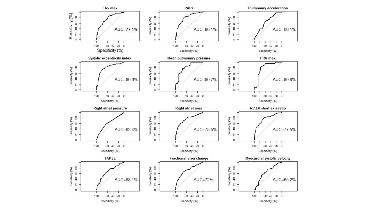
    ScorePermutationsScore
     MinimumMaximum  
    RV systolic pressure mmHg  
     >644857
     >351551
     ≤35 or NA0010
    Right atrial area cm2    
     >253756
     >201241
     ≤20 or NA0010
    Early diastolic pulmonary regurgitation velocity mmHg    
     >364854
     ≥201333
     <20 or NA0010
    RV fractional area change %    
     <351554
     ≥350010
    RV/LV short axis dimension    
     >11663
     ≤1 or NA0000
    Systolic eccentricity index    
     ≥1.11441
     <1.1 or NA0010
    Total permutations n  900 000 
    Maximum score   25

    Each component of the composite score was selected as that having the highest AUC to predict pulmonary hypertension using receiver operating curve analysis (figure 1 and online supplementary material) and consensus. For example, for right ventricular systolic pressure (RVSP), a minimum score of 4 and a maximum of 8 was set; this was done for each threshold and variable, thereby creating a stepwise score. Different combinations of score components (n=900 000) were then tested, and the model with the best AUC chosen, which is displayed as the best score. The order of analysis using the echo composite score is shown in the third column. For example, if a score of 7 was achieved at the first step (if RVSP >64 mmHg), then the echo score became positive to predict severe PH, and no further analysis was needed. If the RVSP was <64 mmHg, the second factor, right atrial area, was considered, and so on. RV: right ventricular; NA: not available; LV: left ventricular.

    • TABLE 2

      Baseline right heart catheter and noninvasive variables

      Total derivation cohortmPAP <25 mmHgmPAP 25–34 mmHgmPAP ≥35 mmHgp-value
      Subjects210467985 
      Age years61±1163±1164±1158±120.004
      Male %555254560.9
      ILD diagnosis  
       CTD59 (28)15 (25)21 (36)23 (39)0.7
       Sarcoidosis43 (20)4 (9)14 (33)25 (58)0.01
       IPF62 (29)18 (29)28 (45)16 (26)0.02
       CHP16 (8)6 (38)2 (12)8 (50)0.1
       NSIP16 (8)2 (12)8 (50)6 (38)0.5
       Other ILD14 (7)1 (7)6 (43)7 (50)0.4
      Right heart catheter  
       mPAP mmHg33±1120±429±343±7<0.001
       PVR Wood units6.0±3.62.6±1.54.6±1.88.8±3.8<0.001
       Cardiac output L·min−1·m−24.3±1.34.8±1.34.1±1.34.1±1.20.02
       PCWP mmHg10±58±510±511±50.008
      BNP ng·L−1102 (44–266)48 (30–72)90 (42–141)241 (105–436)<0.001
      Pulmonary function tests  
       FEV1 L1.6±0.61.6±0.61.5±0.51.6±0.60.9
       FEV1 % pred58±1862±2157±1757±170.2
       FVC L2.0±0.82.0±0.81.9±0.72.2±0.90.2
       FVC % pred60±2061±2259 ±1862±220.7
       TLCO % pred25±1028±1025±924 ±100.04
       KCO % pred52±1759±1854±1648±16<0.001
       PaO2 kPa7.9±1.98.9±1.98.1±1.97.1±1.7<0.001
      CT scan  
       ILD extent <20%/>20%14/8615/8519/819/910.2

      Data are presented as n, mean±sd, n (%) or median (interquartile range), unless otherwise stated. mPAP: mean pulmonary pressure at right heart catheterisation; ILD: interstitial lung disease; CTD: connective tissue disease; IPF idiopathic pulmonary fibrosis; CHP: chronic hypersensitivity pneumonitis; NSIP: nonspecific interstitial pneumonia; PVR: pulmonary vascular resistance; PCWP: pulmonary capillary wedge pressure; BNP: brain natriuretic peptide; FEV1: forced expiratory volume in 1 s; FVC: forced vital capacity; TLCO: transfer factor of the lung for carbon monoxide; KCO: transfer coefficient of the lung for carbon monoxide; PaO2 : arterial oxygen tension (by capillary blood gas analysis); CT: computed tomography.

      • TABLE 3

        Echocardiographic variables grouped according to severity of pulmonary hypertension at right heart catheterisation (RHC)

        Availability %Derivation cohort, totalmPAP <25 mmHgmPAP 25–34 mmHgmPAP ≥35 mmHgp-value
        Subjects 228467985 
        TRVmax m·s−1923.7±0.63.3±0.53.6±0.54.0±0.6<0.001
        RVSP mmHg9266±1953±1361±1876±17<0.001
        Pulmonary acceleration time ms9377±1882±1780±1970±14<0.001
        Systolic eccentricity index821.4±0.41.1±0.21.2±0.31.6±0.5<0.001
        Early PRVmax m·s−1202.5±0.52.0±0.32.3±0.52.7±0.40.001
        RAP mmHg99.55 (5–10)5 (5–10)5 (5–10)10 (5–10)0.008
        Fractional area change %9337±841±839±734±8<0.001
        Right atrial area cm29320±815±418±624±8<0.001
        TAPSE cm921.8±0.51.9±0.41.9±0.51.7±0.4<0.001
        RV:LV short axis dimension ratio (systolic)810.9 (0.7–1.4)0.7 (0.6–0.9)1.0 (0.6–1.1)1.3 (0.9–2.0)<0.001

        Data are presented as n, mean±sd or median (interquartile range), unless otherwise stated. mPAP: mean pulmonary pressure at RHC; TRVmax: maximum tricuspid regurgitation velocity; RVSP: right ventricular systolic pressure; PRVmax: maximum diastolic pulmonary regurgitation velocity; RAP: right atrial pressure; TAPSE: tricuspid annular plane systolic excursion; RV: right ventricular; LV: left ventricular.

        • TABLE 4

          Comparison of the derivation and validation cohorts

          Derivation cohortValidation cohortp-value
          Subjects21061 
          Age years61±1161±130.9
          Male %55390.03
          ILD diagnosis 
           CTD59 (28)33 (54)<0.001
           Sarcoidosis43 (20)6 (10)0.06
           IPF62 (29)5 (8)0.007
           CHP16 (8)6 (10)0.5
           NSIP16 (8)6 (10)0.5
           Other ILD14 (7)5 (8)0.7
          RHC 
           mPAP mmHg33±1133±120.8
           PVR Wood units6.0±3.66.9±5.60.3
           Cardiac output L·min-14.3±1.34.1±1.40.6
           PCWP mmHg10±510±50.9
          BNP ng·L−1102 (44–266)103 (42–306)0.7
          Pulmonary function tests 
           FEV1 L1.6±0.61.6±0.80.5
           FEV1 % pred58±1862±210.3
           FVC L2.0±0.82.0±0.90.7
           FVC % pred60±2065±220.2
           TLCO % pred25±1027±100.2
           KCO % pred52±1754±170.7
           PaO2 kPa7.9±1.98.5±2.10.1
          CT scan 
           ILD extent# 20%/>20%14/8619/810.5

          Data are presented as n, mean±sd or median (interquartile range), unless otherwise stated. ILD: interstital lung disease; CTD: connective tissue disease; IPF idiopathic pulmonary fibrosis; CHP: chronic hypersensitivity pneumonitis; NSIP: nonspecific interstitial pneumonia; RHC: right heart catheterisation; mPAP: mean pulmonary pressure; PVR: pulmonary vascular resistance; PCWP: pulmonary capillary wedge pressure; BNP: brain natriuretic peptide; FEV1: forced expiratory volume in 1 s; FVC: forced vital capacity; TLCO: transfer factor of the lung for carbon monoxide; KCO: transfer coefficient of the lung for carbon monoxide; PaO2: arterial oxygen tension (by capillary blood gas analysis); CT: computed tomography. #: formal scoring of severity on CT (see methods section).

          Supplementary Materials

          • Figures
          • Tables
          • Supplementary Material

            Please note: supplementary material is not edited by the Editorial Office, and is uploaded as it has been supplied by the author.

            Supplementary material 00124-2017_supp

            Figure S1 00124-2017_figureS1

            Figure S2 00124-2017_figureS2

          PreviousNext
          Back to top
          Vol 4 Issue 2 Table of Contents
          ERJ Open Research: 4 (2)
          • Table of Contents
          • Index by author
          Email

          Thank you for your interest in spreading the word on European Respiratory Society .

          NOTE: We only request your email address so that the person you are recommending the page to knows that you wanted them to see it, and that it is not junk mail. We do not capture any email address.

          Enter multiple addresses on separate lines or separate them with commas.
          A stepwise composite echocardiographic score predicts severe pulmonary hypertension in patients with interstitial lung disease
          (Your Name) has sent you a message from European Respiratory Society
          (Your Name) thought you would like to see the European Respiratory Society web site.
          CAPTCHA
          This question is for testing whether or not you are a human visitor and to prevent automated spam submissions.
          Print
          Citation Tools
          A stepwise composite echocardiographic score predicts severe pulmonary hypertension in patients with interstitial lung disease
          Simon Bax, Charlene Bredy, Aleksander Kempny, Konstantinos Dimopoulos, Anand Devaraj, Simon Walsh, Joseph Jacob, Arjun Nair, Maria Kokosi, Gregory Keir, Vasileios Kouranos, Peter M. George, Colm McCabe, Michael Wilde, Athol Wells, Wei Li, Stephen John Wort, Laura C. Price
          ERJ Open Research Apr 2018, 4 (2) 00124-2017; DOI: 10.1183/23120541.00124-2017

          Citation Manager Formats

          • BibTeX
          • Bookends
          • EasyBib
          • EndNote (tagged)
          • EndNote 8 (xml)
          • Medlars
          • Mendeley
          • Papers
          • RefWorks Tagged
          • Ref Manager
          • RIS
          • Zotero
          Share
          A stepwise composite echocardiographic score predicts severe pulmonary hypertension in patients with interstitial lung disease
          Simon Bax, Charlene Bredy, Aleksander Kempny, Konstantinos Dimopoulos, Anand Devaraj, Simon Walsh, Joseph Jacob, Arjun Nair, Maria Kokosi, Gregory Keir, Vasileios Kouranos, Peter M. George, Colm McCabe, Michael Wilde, Athol Wells, Wei Li, Stephen John Wort, Laura C. Price
          ERJ Open Research Apr 2018, 4 (2) 00124-2017; DOI: 10.1183/23120541.00124-2017
          del.icio.us logo Digg logo Reddit logo Technorati logo Twitter logo CiteULike logo Connotea logo Facebook logo Google logo Mendeley logo
          Full Text (PDF)

          Jump To

          • Article
            • Abstract
            • Abstract
            • Introduction
            • Methods
            • Results
            • Discussion
            • Supplementary material
            • Footnotes
            • References
          • Figures & Data
          • Info & Metrics
          • PDF

          Subjects

          • Interstitial and orphan lung disease
          • Tweet Widget
          • Facebook Like
          • Google Plus One

          More in this TOC Section

          Original articles

          • Endobronchial autologous BM-MSCs in IPF patients
          • Effect of β-blockers on the risk of COPD exacerbations
          • Recurrence of symptoms after childhood LRTI
          Show more Original articles

          Interstitial lung disease

          • Electronically monitored medication adherence in IPF
          • ILD incidence and mortality in the UK and the European Union
          • Increased monocytes in early fibrotic ILA
          Show more Interstitial lung disease

          Related Articles

          Navigate

          • Home
          • Current issue
          • Archive

          About ERJ Open Research

          • Editorial board
          • Journal information
          • Press
          • Permissions and reprints
          • Advertising

          The European Respiratory Society

          • Society home
          • myERS
          • Privacy policy
          • Accessibility

          ERS publications

          • European Respiratory Journal
          • ERJ Open Research
          • European Respiratory Review
          • Breathe
          • ERS books online
          • ERS Bookshop

          Help

          • Feedback

          For authors

          • Instructions for authors
          • Publication ethics and malpractice
          • Submit a manuscript

          For readers

          • Alerts
          • Subjects
          • RSS

          Subscriptions

          • Accessing the ERS publications

          Contact us

          European Respiratory Society
          442 Glossop Road
          Sheffield S10 2PX
          United Kingdom
          Tel: +44 114 2672860
          Email: journals@ersnet.org

          ISSN

          Online ISSN: 2312-0541

          Copyright © 2022 by the European Respiratory Society