


Abstract
Synchrotron-based imaging allows for detection of bronchiectasis-like phenotypes in mice with mucociliary clearance disorders https://bit.ly/3gXGdP3
To the Editor:
Bronchiectasis is a chronic pathological condition characterised by abnormal enlargement of the lung's conductive airways. It is associated with a lack of ciliary motility and restricted mucociliary clearance in diseases such as primary ciliary dyskinesia (PCD) or “immotile cilia syndrome”. Recent studies have shown an increase in the prevalence of bronchiectasis, causing a significant burden on public healthcare systems [1, 2]. The mechanisms that trigger and drive the development of bronchiectasis have yet to be fully elucidated. Murine models of immotile cilia or reduced mucociliary clearance failed to display signs of bronchiectasis in multiple studies, raising questions about the suitability of murine models for non-cystic fibrosis (CF) bronchiectasis and hindering the development of targeted therapies [3].
The influence of age and size has been discussed since the duration of mucociliary clearance defects could significantly impact airway remodelling, and many murine models of PCD do not survive long enough for the development of relevant airway remodelling [4]. Moreover, the murine airway anatomy poses significant challenges to the employed imaging modality since most of the conducting airways measure under 1 mm in diameter [5]. Motion artefacts from ventilation and the beating heart further limit the imaging results; they were named the number one technical pitfall in the computed tomography (CT) evaluation of bronchiectasis [6]. Given these limitations, the failure in developing suitable murine models of immotile cilia raises the question: “Could we have simply missed it?”
To answer the question of whether we can detect a phenotype of bronchiectasis or bronchial enlargement under pathogen-free conditions in laboratory mice with mucociliary clearance defects, we generated a cohort of TAp73 wildtype (WT, n=6) and knockout (KO, n=4) mice. All experimental procedures were approved and performed in accordance with the requirements set forth by the Ethics Committee of the University Medical Centre Goettingen (application number: 18/2/16).
TP73, a p53 homologue, has been identified in key steps during the differentiation of motile multiciliated cells and the regulation of airway multiciliogenesis, and TAp73 KO mice have strongly reduced mucociliary clearance [7–9]. Mice were evaluated at 10 months of age (308 days), corresponding to a human age of ∼34 years [10]. At this age, most human patients with severe PCD have already developed visible bronchiectasis on chest CT. Synchrotron-based imaging has been shown to increase structural detectability and permit exact quantification of tissue parameters in small rodents in a variety of applications and organ systems. The high degree of coherence of synchrotron X-ray light allows for phase-sensitive imaging strategies that cannot be applied using commonly available X-ray tubes [11]. One of those is the so-called synchrotron free-propagation phase-contrast CT (SR-pCT), which is characterised by a larger contrast-to-noise ratio in soft-tissue application than classical CT and has been successfully applied for lung imaging in a multitude of studies [12, 13]. The ability of SR-pCT to study the mouse lung in situ in great detail enables reliable depiction of subtle anatomical alterations in their original context [13].
Lessons for clinicians
Murine models of reduced mucociliary clearance have been established to investigate their pathobiology and develop novel treatment approaches but failed to develop visible airway enlargements. Synchrotron free-propagation phase-contrast computed imaging is an innovative, sensitive, nondestructive in situ technique that allows for three-dimensional ultra-high-resolution detection of bronchiectasis in murine models of impaired mucociliary clearance.
Here we utilised SR-pCT to analyse the lung structure at a spatial resolution of ∼9 µm. To maintain the lung architecture, sacrificed mice were tracheotomised and the lungs were inflated with a constant air pressure of 30 cmH2O. The trachea was tied, and the entire mouse was embedded in 1% agarose gel in 50-mL falcon tubes to prevent post mortem deformation or lung deflation [14]. Imaging was performed at the SYRMEP beamline of the Italian synchrotron light source “Elettra” Trieste, Italy using a sample-to-detector distance of 30 cm, a quasi-monochromatic x-ray beam with an energy of 22 keV and a full 360° off-centre scan was performed yielding 1800 angular projections by an effective detector pixel size of 9×9 µm2. Single-distance phase retrieval was applied using the transport of intensity equation [15]. 3D reconstructions were performed utilising a classical filtered back projection algorithm. The delta-to-beta ratio between the decrement of the real part of the complex refractive index and the imaginary part was set to 1950 based on Mohammadi et al. [16]. In the obtained 3D data sets 5–10 volumes-of-interest of a size of 22 mm3 were randomly selected in the periphery of the lung. The average pore size of small conducting airways was then quantified in three dimensions utilising the software “Pore3D”, resulting in a list of pore descriptions for each volume-of-interest. To focus the analysis to smaller bronchi rather than larger airways or parenchymal alveoli, only pores with a size between 0.2 and 0.75 mm were used to assess bronchial dilation. To minimise the influence of differences in the size and weight of the analysed mice we normalised the values with the 3rd root of the weight.
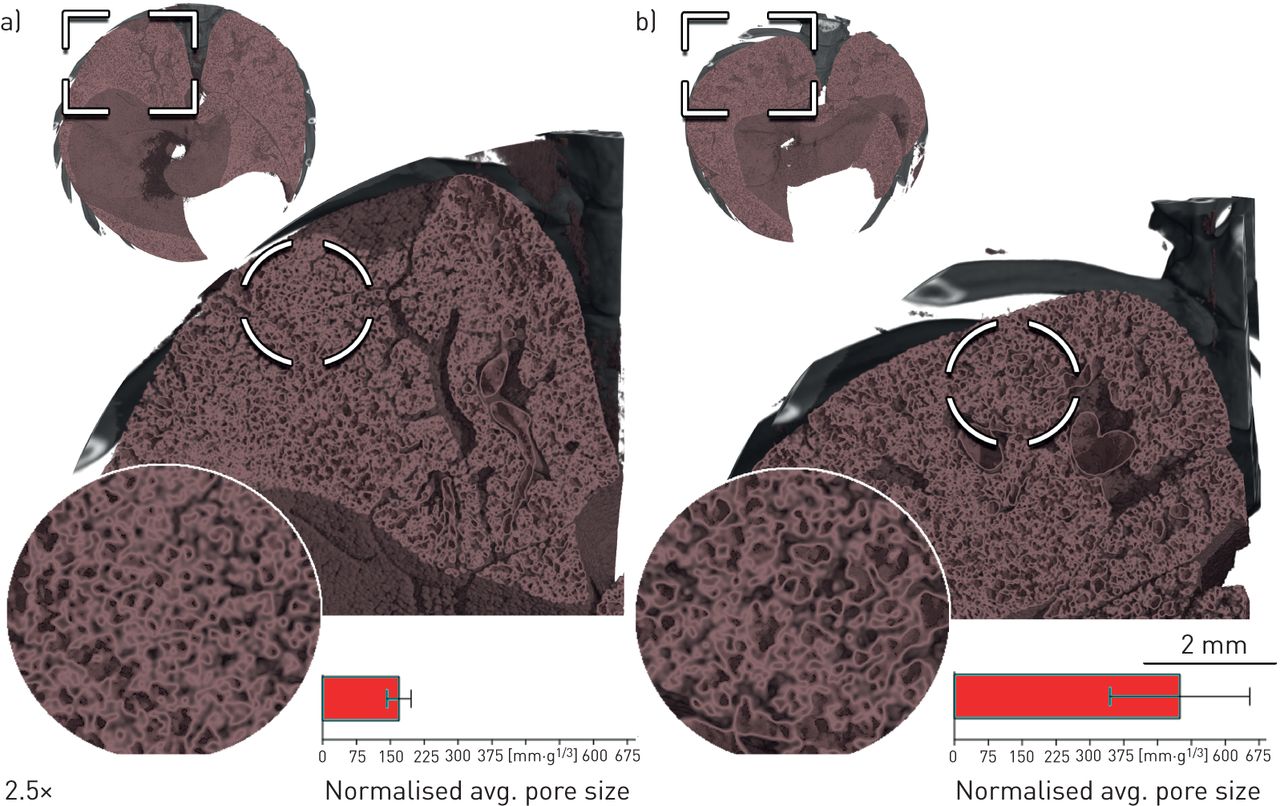
Using SR-pCT we were able to analyse the entire mouse lung in situ on the anatomical level of small conducting airways and found significantly larger normalised airway diameters in the TAp73 KO compared to that found in the WT mice. Side-by-side comparison of a WT control (figure 1a) and a TAp73 KO mouse (figure 1b) shows enlarged small airways in the KO, suggesting the appearance of bronchiectasis-like phenotype in those mice. Statistical evaluation of the cohorts revealed higher porosity of the TAp73 KO lungs with reduced mucociliary clearance.
Synchrotron free-propagation phase-contrast computed tomography in situ analysis (spatial resolution of ∼9 µm) of a) a wildtype (WT) control and b) a TAp73 knockout (KO) mouse with mucociliary clearance defects. Cross-sections of the entire lung are displayed with two levels of magnification of the left lower lobe (p<0.05). The depicted bars present the results for TAp73 KO (n=4) 499±310 mm·g−1/3 and WT (n=6) 169±66 mm·g−1/3 (mean±sd) (p<0.032).
Our study demonstrates that SR-pCT is a sensitive, nondestructive in situ technique for three-dimensional ultra-high-resolution detection of bronchiectasis in mouse models of immotile cilia and restricted mucociliary clearance. Additionally, we show that mice with defective mucociliary clearance do develop measurable airway enlargements at 10 months, corresponding to the mid-thirties in humans. Therefore, SR-pCT may be a useful tool for pre-clinical and translational evaluation of novel treatment strategies and may allow for small rodent research to broaden our understanding of the mechanisms involved in onset and development of airway remodelling and bronchiectasis.
Footnotes
Conflict of interest: W. Wagner has nothing to disclose.
Conflict of interest: C. Dullin has nothing to disclose.
Conflict of interest: S. Andreas reports personal fees from Boehringer Ingelheim and GlaxoSmithKline, and payments for presentations from AstraZeneca, Berlin Chemie, Chiesi and Novartis, outside the submitted work.
Conflict of interest: M. Lizé is an employee of Bayer AG.
- Received September 1, 2020.
- Accepted December 7, 2020.
- Copyright ©ERS 2021
This article is open access and distributed under the terms of the Creative Commons Attribution Non-Commercial Licence 4.0.
References


