Article Text

Abstract
Background Control of asthma is the goal of asthma management worldwide. The Global Initiative for Asthma defined control by a composite measure of clinical findings and future risk but without using markers of airway inflammation, the hallmark of asthma. We investigated whether clinical asthma control reflects eosinophilic inflammation in a broad population.
Methods Control of asthma was assessed over a period of 4 weeks in 111 patients with asthma: 22 totally controlled, 47 well controlled and 42 uncontrolled. Lung function, quality of life, airway hyperresponsiveness to AMP, sputum and blood eosinophils, exhaled nitric oxide (NO) and bronchial biopsies were obtained.
Results The 69 subjects with controlled asthma (totally and well controlled combined) had lower median blood eosinophil numbers, slope of AMP hyperresponsiveness, and alveolar NO levels than the 42 subjects with uncontrolled asthma: 0.18 (range 0.01–0.54) versus 0.22 (0.06–1.16)×109/litre (p<0.05), 3.8 (−0.4–17 750) versus 39.7 (0.4–28 000) mg/ml (p<0.05) and 5.3 (1.5–14.9) versus 6.7 (2.6–51.7) ppb (p<0.05) respectively. Biopsies from subjects with controlled asthma contained fewer eosinophilic granules and more intact epithelium than uncontrolled subjects: 113 (6–1787) versus 219 (19–5313) (p<0.05) and 11.8% (0–65.3) versus 5.6% (0–47.6) (p<0.05) respectively. Controlled asthmatics had better Asthma Quality of Life Questionnaire scores than uncontrolled patients: 6.7 (5.0–7.0) versus 5.9 (3.7–7.0) (p<0.001).
Conclusions The level of asthma control, based on a composite measure of clinical findings, is associated with inflammatory markers, particularly eosinophilic inflammation, with little difference between totally controlled and well controlled asthma.
- Asthma
- Exhaled Airway Markers
- Histology/Cytology
- Asthma Guidelines
Statistics from Altmetric.com
Key messages
-
Control of asthma, based on a composite measure of clinical findings, is associated with direct and indirect markers of inflammation, particularly eosinophils.
-
From the patients’ perspective it is important to note that this composite measure is related to their quality of life.
-
The relationship between clinical and inflammatory measures is not strong, indicating that inflammatory markers provide additional information about a patient's asthma status.
Introduction
Aiming for total control of asthma by applying strict rules for treatment was the focus of the Gaining Optimal Asthma Control (GOAL) study in 2004.1 In this landmark study, totally controlled and well controlled asthma were composite measures based on respiratory symptoms during the day and night, use of rescue medication, peak expiratory flow (PEF) rate, exacerbations, emergency visits to the doctor or hospital, and adverse side effects of treatment.1 Indeed, many patients with uncontrolled asthma improved to a well controlled or totally controlled level after stepwise increase of fluticasone or fluticasone combined with salmeterol. In 2006 the Global Initiative for Asthma (GINA)2 emphasised the importance of asthma control, and also defined it on the basis of a composite measure of clinical indices, with a later modification to include an assessment of the patient's future risk of adverse events. It was recognised that the clinical features of asthma could be well controlled in patients with mild, moderate or severe disease, as indicated by the level of treatment required.3 Surprisingly, a marker of inflammation was not included in the definition of asthma control, whereas chronic airway inflammation is the hallmark of asthma.
Airway inflammation does not correlate well with the level of lung function or symptoms of asthma.4 The question arises as to whether well or totally controlled asthma based on clinical criteria alone sufficiently reflects an adequate control of the underlying airway inflammation. Furthermore, while a patient may not perceive it as important to have minimal airway inflammation, optimal quality of life may be much more important.5 Therefore, a more thorough insight is needed into the complex interplay between clinical control of asthma, quality of life and the underlying airway inflammation. Previous studies have focused on the relationship between asthma severity (defined at the time by symptoms and lung function) and eosinophilic inflammation.5–7 In this observational study we investigated multiple measures of eosinophilic airway inflammation and quality of life in subjects with totally controlled, well controlled and uncontrolled asthma, defined according to the strict GOAL criteria.
Methods
Subjects
Smoking and non-smoking patients with asthma, aged 19–71 years, either using or not using inhaled corticosteroids (ICS), were recruited from research cohorts in Groningen, the Netherlands. All subjects had a past doctor's diagnosis of asthma and documented bronchial hyperresponsiveness to histamine (provocative concentration of histamine causing a 10% fall in forced expiratory volume in 1 s (FEV1) from baseline (PC10 histamine) ≤16 mg/ml, using 30 s tidal breathing). Moreover, patients were included if they had a provocative concentration of AMP causing a 20% fall in FEV1 from baseline (PC20 AMP) ≤320 mg/ml. If PC20 AMP was higher, an additional histamine provocation challenge was performed within 2 weeks and subjects had to demonstrate a PC10 histamine ≤16 mg/ml. The main exclusion criteria were: FEV1 <1.2 litre, chronic obstructive pulmonary disease, bronchiectasis, upper respiratory tract infection (eg, colds) and/or use of antibiotics or oral corticosteroids within 2 months prior to inclusion in the study. The local medical ethics committee approved the study protocol and all subjects gave their written informed consent.
Study design
This prospective cross-sectional study involved subjects paying four visits to the clinic. At visit 1, written informed consent was obtained and patients were enrolled in the study if they fulfilled the inclusion criteria. Patients were instructed how to keep a diary and how to measure PEF at home for the next 4 weeks. At visit 2 (4 weeks later), clinical control of asthma was assessed from the diary according to the GOAL criteria (table E1 in online supplement) and quality of life was determined using the Asthma Quality of Life Questionnaire (AQLQ).8 Blood was drawn for measurement of eosinophils and atopy. Then reversibility of FEV1 was carried out with 800 µg of inhaled albuterol (Ventolin) followed by sputum induction. Within 2 weeks, at visit 3, hyperresponsiveness to AMP was assessed. One to two weeks later this was followed by exhaled nitrogen monoxide (NO) measurements and a bronchoscopy (visit 4).
Atopy
The Phadiatop screening test was used to determine atopic status; it was performed on the ImmunoCap system, according to the manufacturer's instructions (Phadia AB, Uppsala, Sweden). The results are presented as quotients (fluorescence of the serum of interest divided by the fluorescence of a control serum). A positive Phadiatop was defined as patient serum/control serum >1.
Asthma quality of life
The Dutch version of the AQLQ was used for this study.8 The AQLQ is scored on a seven-point scale, with 32 questions on four domains: symptoms, responses to environmental stimuli and the need to avoid them, limitation of activities and emotional dysfunction.
Lung function
FEV1 was measured with a calibrated water-sealed spirometer according to standardised guidelines.9 Reversibility of FEV1% predicted was measured with administration of 800 μg of albuterol. Provocation tests were performed using a method adapted from Cockcroft et al.10 After initial nebulised normal saline, subjects inhaled doubling concentrations of AMP (0.04–320 mg/ml) by 2 min tidal breathing and at 5 min intervals. The slope of the AMP curve was calculated as the ratio between the change in FEV1 (difference between the value at baseline and at final dose of AMP) and the final dose of AMP. Bronchial hyperresponsiveness to histamine was tested as reported previously,11 using the 30 s tidal breathing method and doubling concentrations ranging from 0.13 to 32 mg/ml.
Sputum induction and sputum processing
Sputum was induced by inhalation of hypertonic saline aerosols as previously described.12 Hypertonic saline (5%) was nebulised over three consecutive periods of 5 min. Whole sputum samples were processed according to a method modified from that of Fahy et al.12 ,13 May Grünwald Giemsa staining was used to obtain cell differentials from a total of 600 viable, non-squamous cells. The sputum was not evaluated if the percentage of squamous cells was >80% or if the total number of non-squamous cells was <600.
Alveolar and bronchial nitric oxide
Exhaled NO (eNO) measurement was performed at multiple flow rates (30, 50, 100 and 200 ml/s) on a NIOX (Aerocrine, Stockholm, Sweden). The mean eNO value (ppb) of three technically acceptable attempts per flow rate was used for analysis. The alveolar NO fraction (ppb) and the bronchial NO flux (nl/s) were calculated with a modification of the two-compartment model of NO exchange by Tsoukias and George.14 ,15
Collection, processing, immunohistochemical staining and quantification of bronchial biopsies
After local anaesthesia, bronchial biopsies were obtained using a flexible bronchoscope (Olympus BF P20 or BF XT20) from segmental divisions of the main bronchi. The biopsies were fixed in 4% formalin, processed and embedded in paraffin. Bronchial biopsies were cut into 3 µm thick sections and stained with specific antibodies against a large panel of inflammatory markers. The inflammatory cells were quantified using computer-assisted image analysis (see Methods section in the online supplement for more details).
Statistics
All analyses were performed using SPSS V.14.0.1. The differences in the continuous variables between groups were tested with the Mann–Whitney U test for non-normally distributed data and with the independent sample t test for normally distributed data. PC20 AMP, slope of AMP, blood, sputum and bronchial eosinophils, and eNO were transformed logarithmically to normalise their distribution. We compared the groups' dichotomous variables using the χ2 test and performed multiple logistic regression analysis to investigate which variables were significant determinants for asthma control.
Results
Patient characteristics
One hundred and eleven patients with asthma with a median age of 50 years (range 19–71) were included (table 1). The reasons for not having totally controlled or well controlled asthma are given in the online supplement (table E1). Twenty-two subjects (20%) were totally controlled, 47 (42%) well controlled and 42 (38%) uncontrolled.
Baseline characteristics
Markers of inflammation
The 69 subjects with controlled asthma (totally and well controlled combined) had lower median blood eosinophil numbers than subjects with uncontrolled asthma: 0.18 (range 0.01–0.54) versus 0.22 (0.06–1.16)×109/litre (p<0.05) (figure 1). The slope of PC20 AMP was lower in patients with controlled asthma than in those with uncontrolled asthma: 3.8 (−0.4–17 750) versus 39.7 (0.4–28 000) mg/ml (p<0.05). Alveolar NO levels were lower in subjects with controlled asthma than with uncontrolled asthma: 5.3 (1.5–14.9) versus 6.7 (2.6–51.7) ppb (p<0.05) (Figure 1).

Biological parameters assessed in subjects with asthma. Peripheral blood eosinophils, sputum eosinophils (%), slope of AMP and alveolar nitric oxide concentrations in patients with totally controlled, well controlled and uncontrolled asthma. Each dot represents one subject. Horizontal bars represent median values. No significant differences were found between totally controlled and well controlled asthma. NO, nitric monoxide.
Sputum was evaluable in 72% of the subjects, which may have caused some bias to the results. The total number and proportion of inflammatory cell types did not differ significantly between controlled and uncontrolled subjects (table E2 in online supplement). Post hoc analysis in subgroups of patients with asthma divided according to current smoking status and current corticosteroid use did not affect these results. However, sputum eosinophils were less frequently outside the normal range of >1.9%16 in subjects with controlled asthma versus those with uncontrolled asthma: 9 (19%) versus 13 (39%) respectively (p<0.05). Using a cut-off point >3% values were 7 (16%) versus 12 (36%) respectively (p<0.05). Subjects with controlled asthma had a lower level of eosinophil activation than uncontrolled subjects, as determined by eosinophil peroxidase (EPX) immunopositivity in bronchial biopsies (table 2, figure 2). Controlled asthma was accompanied by an increase in intact epithelium compared with uncontrolled asthma (table 2, figure 2). All other inflammatory and remodelling variables we investigated were comparable in subjects with controlled and uncontrolled asthma (tables 2 and E3 in online supplement).
Inflammation and remodelling in biopsies

Biopsy parameters assessed in subjects with asthma. Eosinophil peroxidase (EPX) immunopositivity (left panel) and the percentage of intact epithelium (right panel) in bronchial biopsies of patients with totally controlled, well controlled and uncontrolled asthma. Each dot represents one subject. Horizontal bars represent median values. No significant differences were found between totally controlled and well controlled asthma.
No differences were found in markers of inflammation between well controlled and totally controlled asthma. However, the slope of PC20 AMP tended to be higher in subjects with totally controlled asthma than in those with well controlled asthma (p=0.08) (figure 1).
Multiple logistic regression analysis on asthma control
Multivariate logistic regression analysis was used to reveal which variables were independently associated with the level of clinical control. Due to multicolinearity all the variables related to eosinophilic inflammation could not be entered simultaneously into the model. We therefore created a model including sex, age, Phadiatop ratio, ICS use and FEV1/vital capacity, in which we entered alveolar NO, blood eosinophils, AMP slope, EPX-positive pixels and the percentage intact epithelium separately. This showed that higher Phadiatop ratio and ICS use were significant determinants of worse asthma control in most of the models. It is noteworthy that a higher AMP slope (OR 0.39; 95% CI 0.24 to 0.63) and higher EPX-positive pixel numbers (OR 0.32; 95% CI 0.13 to 0.79) were significant determinants for worse asthma control (see Results section in online supplement).
Comparison with asthma quality of life
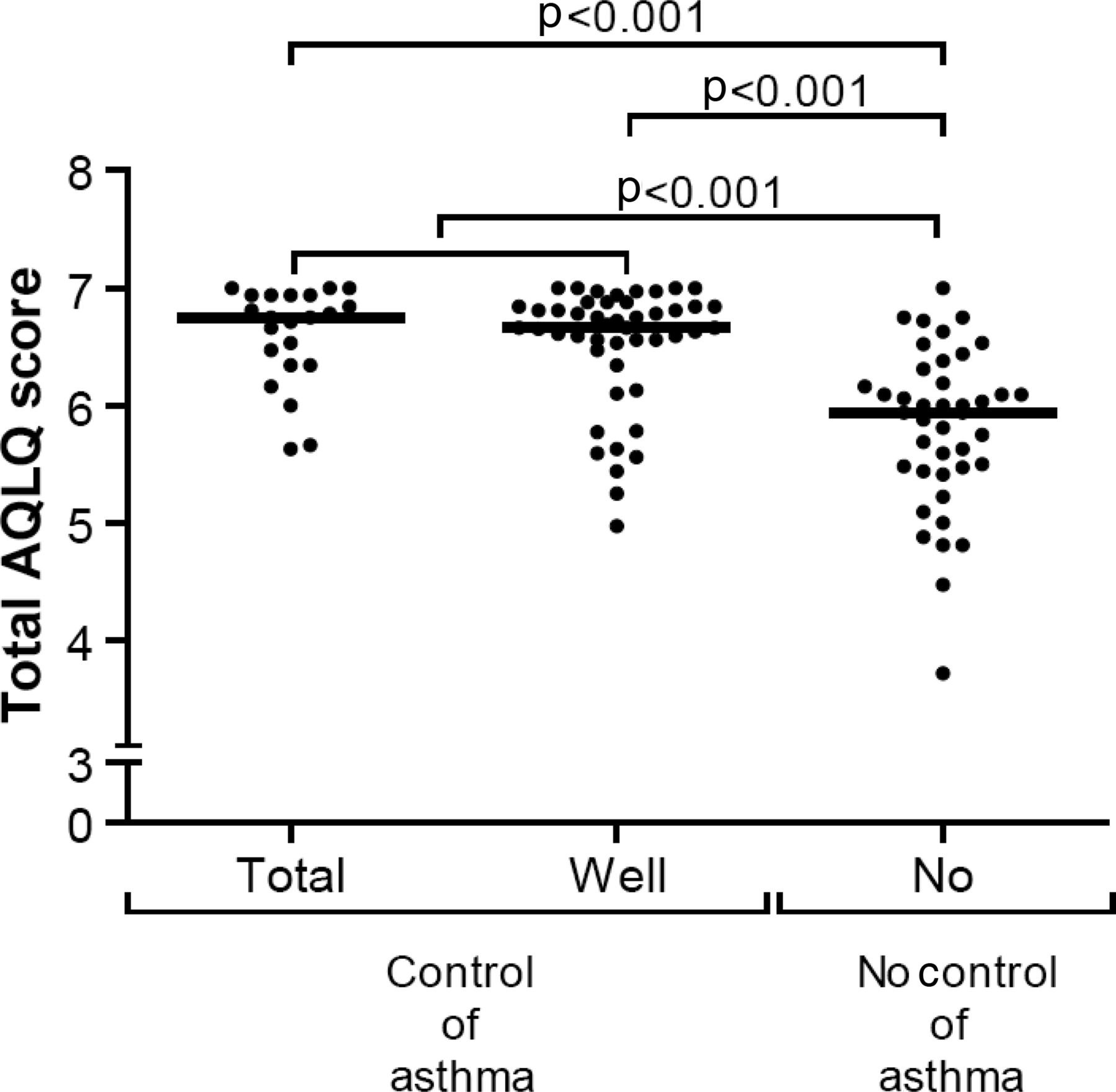
Subjects with controlled asthma demonstrated higher median scores in the total AQLQ than uncontrolled subjects: 6.7 (range 5.0–7.0) versus 5.9 (3.7–7.0) (p<0.001) (figure 3). They also had significantly higher scores in all domains of the AQLQ: symptoms 6.7 (4.9–7.0) versus 5.5 (3.7–7.0), activity 6.8 (4.5–7.0) versus 6.3 (3.9–7.0), emotion 7.0 (5.0–7.0) versus 6.6 (3.8–7.0) and environment 6.5 (3.8–7.0) versus 5.5 (3.0–7.0). AQLQ scores did not differ significantly between subjects with totally controlled and well controlled asthma.

Asthma quality of life as assessed with the Asthma Quality of Life Questionnaire (AQLQ). The AQLQ (Juniper et al8) was given to subjects with totally controlled, well controlled and uncontrolled asthma. It is divided into four domains: symptoms, activities, emotions, environmental. Each dot represents one subject. Horizontal bars represent median values. No significant differences were found between totally controlled and well controlled asthma.
Discussion
The focus of the GINA2 shifted in 2006 from asthma severity before treatment to asthma control during treatment, with assessment based on a composite measure of clinical findings. There is now also considerable interest in controlling not only the clinical manifestations of asthma, but also the inflammatory and pathophysiological features of the disease.16 Although previous studies have described airway inflammation in patients classified by asthma severity, based on past severity classification systems,5–7 our study is the first to investigate whether the level of asthma control according to GOAL is associated with direct and indirect markers of airway inflammation. In a large group of patients with asthma with a wide spectrum of disease severity, patients with totally controlled and well controlled asthma demonstrated less hyperresponsiveness to AMP, lower NO levels in exhaled air, lower eosinophil numbers in peripheral blood, lower activated eosinophil numbers and more epithelial intactness in airway wall biopsies, and higher quality of life scores than subjects with uncontrolled asthma.
Our data suggest that good clinical control of asthma is associated with a lower degree of eosinophilic airway inflammation as assessed by indirect markers like PC20 AMP, eNO and peripheral blood eosinophils. Even more importantly, we found that good control of asthma was associated with less airway inflammation as seen by the classical biopsy findings of mucosal infiltration of activated eosinophils and better epithelial integrity.17 Thus, the concept of a composite measure for clinical asthma control, combining different variables like night-time and daytime symptoms, PEF, and use of rescue medication, appears to have an underlying pathobiological substrate. However, the associations between clinical control and eosinophilic inflammation were not strong, and no significant differences were seen in inflammation markers between totally controlled and well controlled subjects. The lack of correlation between clinical control and eosinophilic inflammation is consistent with emerging evidence for heterogeneity in inflammatory phenotypes, particularly in patients with non-corticosteroid responsive symptoms.18
We further demonstrate that different aspects of inflammation (NO, eosinophils, epithelial cells, PC20 AMP) stemming from different parts of the lung (exhaled air, blood, bronchial biopsies) are associated with good clinical control of asthma. This is not particularly surprising since all these inflammatory factors interact and contribute to the overall, general inflammation that is present in asthma. However, the heterogeneity of asthma with different inflammatory subtypes is increasingly acknowledged.18 ,19 Our study also lends support to this concept, since less severe AMP hyperresponsiveness, lower eosinophil numbers and better epithelial integrity were all independently contributing to better asthma control. Regardless of the exact relationship, our results suggest that patients with better asthma control have less eosinophilic inflammation, although the relationship appeared weaker for sputum eosinophils than for biopsy or blood markers.
We had three unexpected findings in this study. First, the number of pack years smoking or the current smoking status did not contribute significantly to the level of asthma control, despite the fact that smoking has been associated with poorer asthma control in other studies.20–22 One explanation could be that the number of pack years smoking was low, just as the proportion of current smokers. Another explanation might be a ‘healthy smoker’ effect, that is, those with relatively healthy airways are able to tolerate smoke inhalation, to persist in their smoking habit while still showing good control of their asthma.
Second, ICS use contributed to the variation in asthma control, but in an opposite direction to that intuitively expected, that is, patients with uncontrolled asthma used ICS more frequently and with higher doses than patients with controlled asthma. This is likely to represent indication bias, in that patients with worse asthma control are more likely to be prescribed ICS. In addition, an ‘unhealthy ICS’ effect may have also been present in our study since it is known that some patients with asthma do not respond well to corticosteroids.23 ,24 This was also found in the GOAL study, in which 20–30% of the participants were still classified as having uncontrolled asthma at the end of the study despite long-term administration of increasing doses of ICS.
Third, the level of asthma control by GOAL criteria did not significantly associate with lower sputum eosinophil levels, although significantly more patients with uncontrolled asthma had sputum eosinophils outside the normal range.16 Three previous studies, using a range of criteria for asthma control, have shown different results. Romagnoli et al25 (n=35) found median sputum eosinophil levels of 0% and 2.5% (p=0.01) in patients classified as having controlled and poorly controlled asthma, based on symptom frequency, night awakening, bronchodilator use and PEF variability. Quaedvlieg et al26 (n=134) reported median sputum eosinophil levels of 0.4%, 1.4% and 5.6% (p=0.001 well controlled vs uncontrolled) for patients with asthma classified as well controlled, borderline and uncontrolled respectively by the six-item Asthma Control Questionnaire.27 In contrast, Shiota et al28 (n=96) found no significant difference in sputum eosinophil levels for patients classified as having total, partial and uncontrolled asthma by the Asthma Control Test29 (2.1%, 3.8% and 4.9% respectively, p=0.4). The differing eosinophil levels seen in these studies emphasise the potential variation due to patient selection. Unlike earlier studies, the present study also included eosinophilic markers in peripheral blood and airway wall biopsies, and these were found to better reflect a strict definition of asthma control. It is an intriguing finding that control of asthma significantly associates with lower peripheral blood eosinophils and lower activated eosinophil numbers in the bronchial wall, yet only weakly with sputum eosinophils. Nevertheless, across all three compartments, the findings demonstrate that assessment of airway inflammation provides additional information about asthma status than is obtained from clinical control measures.
The present study also confirms that health-related quality of life in patients with controlled asthma is significantly better than in uncontrolled asthma. As the difference between the two groups was higher than the minimal, clinically important difference of 0.5 point, this emphasises the overall impact of asthma control on health status.30 In addition, as in the GOAL study itself,31 little difference was seen in absolute AQLQ scores between subjects with well controlled and totally controlled asthma, suggesting that further improvement in clinical asthma control may not be reflected in a clinically important difference in an individual's quality of life.
The participants in this study are not fully comparable to those in the larger GOAL study,1 ,32 which included only subjects with uncontrolled asthma with a smoking history of <10 pack years, whereas we also included subjects with totally controlled and well controlled asthma with no limit on smoking history. The GOAL study also included younger subjects (including children) and had a lower prevalence of ex-smokers, had lower lung function and higher bronchodilator reversibility. When comparing the reasons why patients failed to achieve total asthma control at baseline in the GOAL study32 and in our study (Table E1) there are a few striking differences. The percentages of patients failing to achieve total control due to the GOAL criteria were 63% due to awakening in GOAL versus 19% in our study, daytime symptoms (95% vs 34%), rescue medication (92% vs 26%), and PEF ≥80% predicted (72% vs 50%). Thus, the prevalence of daytime symptoms and rescue medication in the GOAL study were considerably higher than in our study. It is possible that the patients in the GOAL study had more severe disease because they also had a lower baseline lung function and more symptoms than our patients. Larger, explorative studies, also including patients with more severe asthma, will be needed to determine why some patients do not achieve total control of their asthma and this may have important consequences for the therapies prescribed.
In conclusion, our study demonstrates that clinical control of asthma is associated with direct and indirect markers of airway inflammation, but that airway inflammation provides different information about asthma status from that provided by clinical measures of control. Moreover, better asthma control associates with a higher quality of life for the patients. Although aiming for good clinical control of asthma, as recommended by the GINA guidelines, is important for suppressing the underlying airway inflammation and from a patient's perspective, the present findings do not support a routine increase in ICS treatment for patients with well controlled asthma. Longitudinal intervention studies are needed to assess if the control of asthma concept may recognise patients with asthma who are at higher future risk of exacerbations, accelerated decline in lung function and side effects of treatment.
Acknowledgments
The authors would like to thank all the participants and the lung function department of Beatrixoord, Groningen for their help in collecting all the lung function and sputum data. We thank Dr J Vonk for help with the final statistical analysis and J Senior for critically reading the manuscript. We thank Dr J Lee and Dr N Lee (Mayo Clinic, Scottsdale, Arizona, USA) for providing the anti-EPX antibody.
References
Supplementary materials
Supplementary Data
This web only file has been produced by the BMJ Publishing Group from an electronic file supplied by the author(s) and has not been edited for content.
Files in this Data Supplement:
- Data supplement 1 - Online supplement
Footnotes
-
Funding GlaxoSmithKline gave an unrestricted grant (SAM101761) to this investigator initiated study.
-
Contributors DSP, WT, MNH, and NHTtH were all involved in the design of the study, carrying-out the study, analysis of data, interpretation of results, and reviewing of the manuscript. FV recruited the patients and coordinated the patient-related investigations and organised a database. MEL performed all lab work, including the quantitate analysis of biopsies and sputum. MB was also involved in the quantitative analysis of biopsies and sputum; additionally she did the statistical analyses and wrote the first drafts of the manuscript together with NHTtH. HKR reviewed the manuscript and contributed to some major changes. FV and MB contributed equally.
-
Competing interests Professor Postma consulted to AstraZeneca, Boehringer Ingelheim, GSK, Nycomed and Teva, and received grants from GlaxoSmithKline, Chiesi, Nycomed and AstraZeneca. Dr ten Hacken received grants from Chiesi, GlaxoSmithKline, Nycomed, Boehringer Ingelheim. Dr Reddel has provided consulting for AstraZeneca and Novartis, is on a data monitoring committee for AstraZeneca, GlaxoSmithKline, Merck and Novartis, and has received grants from GlaxoSmithKline. The other authors have no competing interests.
-
Ethics approval Ethical Committee of the University Medical Center Groningen.
-
Provenance and peer review Not commissioned; externally peer reviewed.
Linked Articles
- Airwaves